
As your baby grows steadily throughout pregnancy, they usually move into a head-down position during the third trimester. Since the head is the heaviest part of the body, this position is considered the ideal posture for a vaginal birth.
However, for various reasons, some babies do not move into the head-down position. This condition is known as a breech presentation.
The following facts are known about breech babies.
Approximately 20–30% of fetuses are in a breech position between 24 and 27 weeks of pregnancy.
Most babies naturally turn into the head-down position by around 28 weeks. However, even after turning, some babies may return to the breech position.
Most pregnant women do not notice that their baby is breech because it usually causes no obvious symptoms. In most cases, the condition is first identified during a routine prenatal checkup.
It is also common for doctors to recommend simply monitoring the situation. However, if the baby is still breech at around 33–34 weeks of pregnancy, a scheduled Cesarean section (C-section) may suddenly be recommended. Many expectant mothers only realize at that point that a vaginal delivery may no longer be possible.
Prenatal checkups are generally performed every four weeks until 23 weeks of pregnancy and every two weeks from 24 weeks onward. Therefore, it is usually impossible to determine exactly when the baby first became breech.
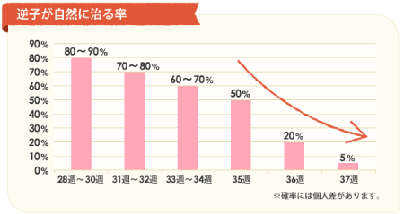
Approximately 3–5% of babies remain in the breech position until delivery. For the safety of both mother and baby, a Cesarean section is recommended in most of these cases.
Even if a breech presentation is diagnosed between 24 and 27 weeks of pregnancy, many babies naturally turn head-down, so expectant mothers are often told not to worry too much.
Previously, we recommended starting our Breech Baby Improvement Program around 28 weeks because babies are still relatively small and can turn more easily. After 34 weeks, babies become larger, making it more difficult for them to rotate naturally.

We have also found that the longer a baby remains in the breech position, the more difficult it may become to correct.
At Medical Japan, we recommend starting our
Breech Baby Improvement Program as early as possible after a breech diagnosis
to help encourage the baby to turn naturally and reduce the likelihood of returning to the breech position.
Although commonly referred to simply as “breech,” there are several different fetal positions. They are generally classified into the following three categories.
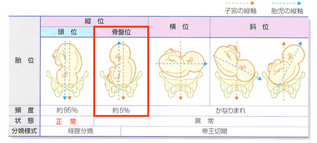
The baby is positioned diagonally within the uterus. This is also quite rare.
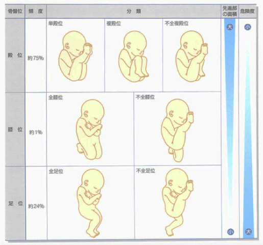
If you are told during a prenatal examination that your baby is breech, ask your healthcare provider where the baby’s head and feet are positioned. This can help you understand changes over time.
Even if the baby remains breech at the next examination, a different position may indicate that the baby is still moving freely and may turn naturally. If the position has not changed at all, the breech presentation may be more difficult to correct.
The exact cause of breech presentation is not fully understood. However, several factors are believed to increase the likelihood of a baby remaining in the breech position.
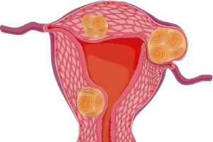

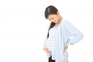
Abnormalities in the shape or position of the uterus or placenta may restrict the baby’s movement within the uterus, increasing the likelihood of a breech presentation.
• Uterine abnormalities
• Uterine fibroids
• Contracted pelvis (narrow pelvis)
• Placental abnormalities (placenta previa, low-lying placenta), etc.
Certain conditions affecting the baby or the uterine environment may limit the baby’s ability to move freely, increasing the likelihood of a breech presentation.
• Polyhydramnios (excess amniotic fluid)
• Multiple pregnancy (twins or more)
• Extended fetal legs
• Low birth weight fetus
• Fetal abnormalities (such as anencephaly or hydrocephalus), etc.
The following lifestyle factors may reduce blood flow to the uterus. As a result, the baby may have less room or ability to move freely, increasing the likelihood of remaining in a breech position.
• Poor posture (rounded shoulders/slouching)
• Rapid weight gain
• Lack of physical activity
• Stress
• Sleep deprivation
• Coldness in the legs and lower back
• Fatigue, etc.
A breech presentation may be associated with the following risks and complications.
In footling or kneeling breech presentations, the baby’s leg movements may cause premature rupture of the membranes before the due date. This may lead to umbilical cord prolapse or even fetal prolapse, both of which are serious obstetric emergencies that can threaten the baby’s life.
During a normal delivery, the baby’s head—the largest part of the body—helps widen the birth canal. However, in a vaginal breech delivery, the baby’s arms or legs may become trapped, increasing the risk of shoulder or hip dislocation. Since the head is delivered last, labor can become significantly more difficult.
Reduced oxygen supply to the baby’s brain during delivery may result in birth asphyxia, neurological complications, or, in rare cases, fetal death.
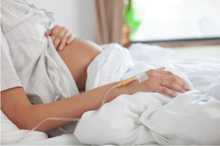
From the perspective of modern obstetrics, a planned Cesarean section is generally considered the safest option for both mother and baby when the baby remains in a breech position. Because there is no universally effective medical treatment to correct breech presentation, and due to safety considerations, most breech pregnancies today are delivered by planned Cesarean section.
Although the timing is determined by the attending obstetrician, a planned Cesarean section is generally scheduled at around
37–38 weeks of pregnancy,
taking the following factors into consideration.
• Delivery is scheduled before labor begins or the membranes rupture naturally, reducing potential risks.
During a vaginal breech delivery, the baby’s head—the largest part of the body—is delivered last, increasing the risk of oxygen deprivation. The smaller the presenting part (for example, a foot compared with the buttocks), the less the birth canal is able to dilate, making delivery more dangerous. The risk generally increases in the following order: frank breech → kneeling breech → footling breech.
In addition, traction on the baby’s arms or legs during delivery may increase the risk of joint injuries such as dislocation. For these reasons, a planned Cesarean section is generally recommended for most breech pregnancies.


At Medical Japan, treatment is performed while continuously monitoring the baby using ultrasound imaging. This approach allows us to minimize potential risks and provide treatment with a strong emphasis on safety.
“`html
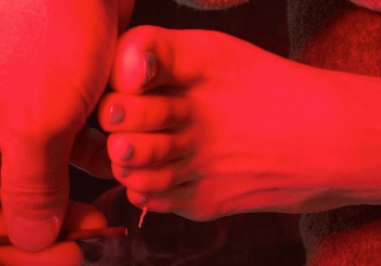

As part of our approach to breech presentation, moxibustion is applied to Sanyinjiao, located above the inner ankle, and Zhiyin, located near the little toe. We offer different types of moxibustion according to each patient’s constitution and sensitivity, ranging from smokeless electric warming devices designed to minimize the risk of burns to traditional moxibustion that produces smoke.
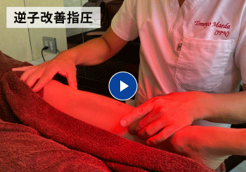
Based on many years of clinical experience, acupressure applied to specialized points may provide effective stimulation. This approach utilizes the somatovisceral reflex, in which stimulation applied to the surface of the body may influence responses in the internal organs.
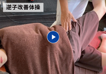
In addition, exercise therapy such as Pilates can help improve pelvic alignment, create a more comfortable environment for the baby, and reduce the risk of recurrence. Our independently developed “4D Exercise” program is particularly suitable for improving the pelvic environment.
This allows us to select acupuncture, moxibustion, acupressure, or a combination of techniques and provide the most appropriate level of stimulation for each patient.
Improvements may still be seen in patients who are
more than 32 weeks pregnant,
so please feel free to contact us for a consultation.

32-year-old woman living in Tachikawa City
She visited our center after her baby moved into a breech position at 30 weeks of pregnancy.
Full-body alignment adjustment
Moxibustion applied to the legs at Sanyinjiao
Acupressure applied to specialized points on the feet
She continued to attend the center for recurrence prevention and general maternity care. The breech presentation did not recur, and she safely delivered a healthy baby boy.
*Breech presentation may recur depending on its underlying causes. For this reason, our center recommends continued care.

The Role of Preconception Care Throughout the Life Course
(Quoted and adapted from the
WHO)
“Conception” means becoming pregnant and welcoming a new life into the womb.
Preconception care refers to the process by which women and couples consider their lifestyle and health in preparation for a possible future pregnancy.
This care is not limited to women who are currently planning a pregnancy. Its goals are to help women and couples become healthier, increase their chances of having a healthy baby, and support healthier lives for women and their future families.
In Japan, the first Preconception Care Center was established at the National Center for Child Health and Development.
The Preconception Care Center aims to provide all women and couples who may wish to have children in the future with comprehensive assessments of their current health and opportunities to consult specialists from different fields about their daily lifestyle and health.
Particular attention is given to consultations about future pregnancy and childbirth for people with current medical conditions or illnesses experienced during childhood, couples who are having difficulty conceiving, and couples who have not achieved their desired outcome in previous pregnancies.

The center also collects information about pregnancy and childbirth involving medical conditions for which sufficient pregnancy-related information has not previously been available. It provides up-to-date information that everyone should know before becoming pregnant.
• Department of Obstetrics and Gynecology, The Jikei University School of Medicine
• Kyorin University Hospital
• Osaka Medical and Pharmaceutical University Hospital
*These facilities provide preconception consultation services only.
“`
© Copyright (C) All Rights Reserved..